Unveiling The Secrets: The Driving Force Behind Chromatid Movement In Mitosis
During cell division, the replicated chromosomes condense and become visible as chromatids. These chromatids must be separated and moved to opposite poles of the cell in order for cell division to be successful. The structure responsible for moving the chromatids is the spindle apparatus.
The spindle apparatus is a complex structure composed of microtubules, motor proteins, and other proteins. Microtubules are long, thin fibers that extend from one pole of the cell to the other. Motor proteins are proteins that can walk along microtubules, carrying chromatids with them. The spindle apparatus is responsible for separating the chromatids and moving them to opposite poles of the cell.
The spindle apparatus is essential for cell division. Without it, the chromatids would not be able to separate and move to opposite poles of the cell, and cell division would not be able to occur. The spindle apparatus is a complex and dynamic structure that plays a vital role in cell division.
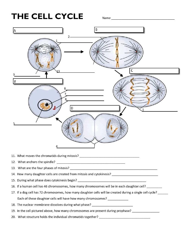
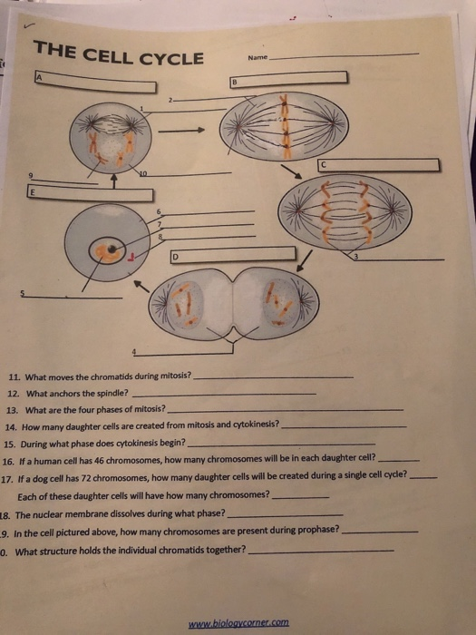
What Moves the Chromatids During Mitosis
During cell division, the replicated chromosomes condense and become visible as chromatids. These chromatids must be separated and moved to opposite poles of the cell in order for cell division to be successful. The structure responsible for moving the chromatids is the spindle apparatus.
- Spindle apparatus: The structure that moves the chromatids.
- Microtubules: Long, thin fibers that make up the spindle apparatus.
- Motor proteins: Proteins that walk along microtubules, carrying chromatids with them.
- Kinetochore: The protein complex that attaches the spindle apparatus to the chromatids.
- Centromere: The region of the chromosome where the kinetochore is located.
- Metaphase plate: The plane in the center of the cell where the chromosomes line up before they are separated.
- Anaphase: The stage of mitosis when the chromatids are separated and moved to opposite poles of the cell.
- Telophase: The stage of mitosis when the chromosomes reach the poles of the cell and the spindle apparatus disappears.
- Cytokinesis: The division of the cytoplasm that follows mitosis.
- Cell division: The process of dividing a cell into two new cells.
These key aspects are all essential for understanding how the chromatids are moved during mitosis. The spindle apparatus is the structure that physically moves the chromatids, while the microtubules and motor proteins provide the force necessary for movement. The kinetochore is the point of attachment between the spindle apparatus and the chromatids, and the centromere is the region of the chromosome where the kinetochore is located. The metaphase plate is the plane in the center of the cell where the chromosomes line up before they are separated, and anaphase is the stage of mitosis when the chromatids are separated and moved to opposite poles of the cell. Telophase is the stage of mitosis when the chromosomes reach the poles of the cell and the spindle apparatus disappears, and cytokinesis is the division of the cytoplasm that follows mitosis.
Spindle apparatus
The spindle apparatus is a complex structure composed of microtubules, motor proteins, and other proteins. Microtubules are long, thin fibers that extend from one pole of the cell to the other. Motor proteins are proteins that can walk along microtubules, carrying chromatids with them. The spindle apparatus is responsible for separating the chromatids and moving them to opposite poles of the cell.
The spindle apparatus is essential for cell division. Without it, the chromatids would not be able to separate and move to opposite poles of the cell, and cell division would not be able to occur. The spindle apparatus is a complex and dynamic structure that plays a vital role in cell division.
One of the most important functions of the spindle apparatus is to ensure that the chromatids are separated equally. This is essential for the proper development of the new cells. If the chromatids are not separated equally, it can lead to genetic abnormalities, such as Down syndrome.
The spindle apparatus is also involved in the process of cytokinesis, which is the division of the cytoplasm. The spindle apparatus helps to position the chromosomes in the center of the cell, and it also helps to form the cleavage furrow, which is the indentation in the cell membrane that eventually divides the cell into two new cells.
The spindle apparatus is a complex and essential structure that plays a vital role in cell division. It is responsible for separating the chromatids and moving them to opposite poles of the cell, and it also helps to position the chromosomes in the center of the cell and form the cleavage furrow.
Microtubules
Microtubules are long, thin fibers that make up the spindle apparatus, which is the structure that moves the chromatids during mitosis. Microtubules are composed of a protein called tubulin, and they are organized into a hollow cylinder. The spindle apparatus is composed of two sets of microtubules, which are arranged in a bipolar fashion. The poles of the spindle apparatus are located at opposite ends of the cell, and the microtubules extend from each pole to the center of the cell.
During mitosis, the spindle apparatus is responsible for separating the chromatids and moving them to opposite poles of the cell. The microtubules of the spindle apparatus attach to the kinetochores, which are protein complexes located at the centromeres of the chromosomes. Once the microtubules are attached to the kinetochores, the motor proteins dynein and kinesin move along the microtubules, pulling the chromatids to opposite poles of the cell. This process is known as chromosome segregation.
Microtubules are essential for the proper segregation of chromosomes during mitosis. If the microtubules are not functioning properly, the chromosomes will not be able to separate properly, and this can lead to cell death. Microtubules are also involved in other cellular processes, such as cell division, cell migration, and organelle transport.
The study of microtubules has led to the development of new drugs that target microtubules. These drugs are used to treat a variety of diseases, including cancer and neurodegenerative diseases.
Motor proteins
Motor proteins are essential for moving the chromatids during mitosis. They are proteins that can walk along microtubules, carrying chromatids with them. Motor proteins use the energy from ATP to move along the microtubules, and they can move in either direction along the microtubule.
During mitosis, motor proteins attach to the kinetochores of the chromosomes. Kinetochores are protein complexes located at the centromeres of the chromosomes. Once the motor proteins are attached to the kinetochores, they begin to move along the microtubules, pulling the chromatids with them. This process is known as chromosome segregation.
Motor proteins are essential for the proper segregation of chromosomes during mitosis. If the motor proteins are not functioning properly, the chromosomes will not be able to separate properly, and this can lead to cell death. Motor proteins are also involved in other cellular processes, such as cell division, cell migration, and organelle transport.
The study of motor proteins has led to the development of new drugs that target motor proteins. These drugs are used to treat a variety of diseases, including cancer and neurodegenerative diseases.
Kinetochore
The kinetochore is a protein complex that attaches the spindle apparatus to the chromatids. It is located at the centromere of the chromosome, which is the narrow region of the chromosome where the two sister chromatids are joined. The kinetochore is responsible for ensuring that the chromatids are properly attached to the spindle apparatus and that they are pulled apart correctly during cell division.
- Structure of the kinetochore
The kinetochore is a complex structure that is composed of several different proteins. These proteins interact with the microtubules of the spindle apparatus and with the DNA of the chromosome. The kinetochore is also the site of attachment for the motor proteins that pull the chromatids apart during cell division.
- Function of the kinetochore
The kinetochore plays a vital role in cell division. It ensures that the chromatids are properly attached to the spindle apparatus and that they are pulled apart correctly. This is essential for the proper segregation of the chromosomes during cell division.
- Regulation of the kinetochore
The kinetochore is regulated by a number of different factors, including the cell cycle checkpoints. These checkpoints ensure that the kinetochore is properly attached to the spindle apparatus before cell division proceeds. The kinetochore is also regulated by a number of different proteins, which help to ensure that the kinetochore functions properly.
- Errors in kinetochore function
Errors in kinetochore function can lead to a number of different problems, including chromosome missegregation. Chromosome missegregation can lead to a number of different genetic disorders, including Down syndrome and cancer.
The kinetochore is a complex and essential structure that plays a vital role in cell division. It ensures that the chromatids are properly attached to the spindle apparatus and that they are pulled apart correctly. This is essential for the proper segregation of the chromosomes during cell division.
Centromere
The centromere is a specialized region of the chromosome that is essential for chromosome segregation during cell division. It is the site of attachment for the mitotic spindle fibers, which are responsible for pulling the chromosomes to opposite poles of the cell. The kinetochore, a large protein complex that assembles at the centromere, provides the physical link between the chromosomes and the mitotic spindle.
The centromere is composed of highly repetitive DNA sequences that do not code for any proteins. These repetitive sequences serve as binding sites for kinetochore proteins, which are essential for the proper attachment of the chromosomes to the mitotic spindle. The kinetochore is a complex structure that consists of more than 100 different proteins, which work together to ensure that the chromosomes are properly attached to the spindle fibers and that they are pulled apart correctly during cell division.
The centromere is essential for the proper segregation of chromosomes during cell division. If the centromere is not properly attached to the mitotic spindle, the chromosomes will not be pulled apart correctly, and this can lead to aneuploidy, which is a condition in which a cell has an abnormal number of chromosomes. Aneuploidy is a common cause of birth defects and developmental disorders, such as Down syndrome.
The study of the centromere is important for understanding the fundamental mechanisms of cell division. It is also important for understanding the causes of aneuploidy and developing new treatments for aneuploidy-related diseases.
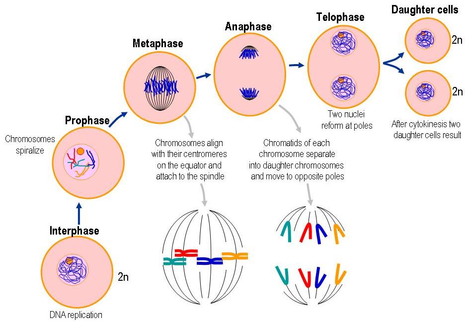
Metaphase plate
The metaphase plate is a crucial component of the process of cell division, particularly during mitosis. It serves as the central platform where all chromosomes are aligned and organized prior to their separation and movement to opposite poles of the cell. This alignment ensures that each daughter cell receives an equal complement of genetic material.
The formation of the metaphase plate is facilitated by the mitotic spindle, a complex structure composed of microtubules and motor proteins. These microtubules extend from opposite poles of the cell and attach to the chromosomes at specialized regions called kinetochores. The kinetochores are located at the centromere of each chromosome, which is the narrow region where sister chromatids are joined.
Once the chromosomes are attached to the microtubules of the mitotic spindle, they are pulled towards the center of the cell. This movement is driven by the motor proteins, which use the energy from ATP to walk along the microtubules. As the chromosomes move, they become aligned along the metaphase plate, ensuring that they are positioned equidistantly from the two poles of the cell.
The alignment of chromosomes on the metaphase plate is essential for the accurate segregation of genetic material during cell division. If the chromosomes are not properly aligned, they may not be separated equally between the two daughter cells, leading to genetic abnormalities. Therefore, the metaphase plate plays a critical role in maintaining the stability and integrity of the genome.
Anaphase
Anaphase is a crucial stage of mitosis, the process by which a cell divides into two identical daughter cells. During anaphase, the chromatids, which are identical copies of each chromosome, are separated and moved to opposite poles of the cell. This movement is driven by the mitotic spindle, a complex structure composed of microtubules and motor proteins.
- Role of the mitotic spindle
The mitotic spindle is responsible for separating the chromatids and moving them to opposite poles of the cell. The mitotic spindle is composed of microtubules, which are long, thin fibers that extend from one pole of the cell to the other. Motor proteins, which use energy from ATP to walk along microtubules, attach to the kinetochores of the chromosomes and pull them apart.
- Kinetochores
Kinetochores are protein complexes that attach the chromosomes to the mitotic spindle. Each kinetochore is located at the centromere of a chromosome, which is the narrow region where sister chromatids are joined. The kinetochores ensure that the chromosomes are properly attached to the mitotic spindle and that they are pulled apart correctly during anaphase.
- Errors in anaphase
Errors in anaphase can lead to aneuploidy, a condition in which a cell has an abnormal number of chromosomes. Aneuploidy can cause a variety of genetic disorders, including Down syndrome and cancer.
Anaphase is a critical stage of mitosis, and it is essential for the proper segregation of chromosomes. Errors in anaphase can lead to aneuploidy, which can cause a variety of genetic disorders.
Telophase
Telophase is the final stage of mitosis, the process by which a cell divides into two identical daughter cells. During telophase, the chromosomes, which are identical copies of each chromosome, reach the poles of the cell and the spindle apparatus disappears.
- Role of telophase in chromosome segregation
Telophase plays a critical role in chromosome segregation, the process by which the chromosomes are separated and distributed to the two daughter cells. The spindle apparatus, which is responsible for separating the chromosomes, disassembles during telophase, allowing the chromosomes to reach the poles of the cell.
- Nuclear envelope reformation
During telophase, the nuclear envelope, which surrounds the nucleus, reforms around each set of chromosomes. The nuclear envelope is a double membrane that protects the chromosomes and regulates the movement of materials into and out of the nucleus.
- Cytokinesis
Telophase is followed by cytokinesis, the process by which the cytoplasm of the cell is divided into two daughter cells. Cytokinesis can occur by two different mechanisms: pinching in the middle (animal cells) or forming a cell plate (plant cells).
- Completion of mitosis
Telophase marks the completion of mitosis. The two daughter cells are now independent and can enter the next phase of the cell cycle, which is interphase.
Telophase is a critical stage of mitosis, and it is essential for the proper segregation of chromosomes. Errors in telophase can lead to aneuploidy, a condition in which a cell has an abnormal number of chromosomes. Aneuploidy can cause a variety of genetic disorders, including Down syndrome and cancer.
Cytokinesis
Cytokinesis is the division of the cytoplasm that follows mitosis. It is the final step in cell division, and it results in the formation of two daughter cells. Cytokinesis is essential for the proper segregation of the chromosomes, as it ensures that each daughter cell receives an equal complement of genetic material.
There are two main types of cytokinesis: pinching in the middle (animal cells) and forming a cell plate (plant cells). In animal cells, cytokinesis occurs when a cleavage furrow forms around the equator of the cell. The cleavage furrow is a shallow groove that is caused by the constriction of the actin filaments in the cell cortex. As the cleavage furrow grows deeper, it eventually pinches the cell in two, forming two daughter cells.
In plant cells, cytokinesis occurs when a cell plate forms in the center of the cell. The cell plate is a new cell wall that is formed by the fusion of vesicles from the Golgi apparatus. As the cell plate grows, it eventually divides the cell into two daughter cells.
Cytokinesis is an essential part of cell division. It ensures that each daughter cell receives an equal complement of genetic material, and it prevents the formation of multinucleated cells.
Cell division
Cell division is the process by which a cell divides into two new cells. It is a fundamental process in biology, as it is essential for growth, development, and reproduction. There are two main types of cell division: mitosis and meiosis.
Mitosis is the process by which a cell divides into two identical daughter cells. It is used for growth and development. Meiosis is the process by which a cell divides into four haploid daughter cells. It is used for reproduction.
During mitosis, the chromosomes in the cell condense and become visible. The spindle apparatus, which is made up of microtubules, forms and attaches to the chromosomes. The chromosomes are then pulled apart by the spindle apparatus and moved to opposite poles of the cell. Once the chromosomes are at the poles of the cell, the spindle apparatus disappears and the nuclear envelope reforms around each set of chromosomes. The cell then divides into two daughter cells.
The movement of the chromosomes during mitosis is essential for the proper segregation of the genetic material. If the chromosomes are not properly segregated, it can lead to genetic abnormalities, such as Down syndrome.
Cell division is a complex and essential process in biology. It is essential for growth, development, and reproduction. The movement of the chromosomes during mitosis is a critical step in cell division, and it is essential for the proper segregation of the genetic material.
FAQs about "What Moves the Chromatids During Mitosis"
The movement of chromatids during mitosis is a crucial process in cell division, ensuring the equal distribution of genetic material to daughter cells. Here are some frequently asked questions to address common concerns and misconceptions about this topic:
Question 1: What is the structure responsible for moving chromatids during mitosis?
Answer: The spindle apparatus, composed of microtubules, motor proteins, and other proteins, is responsible for the movement and separation of chromatids during mitosis.
Question 2: How do motor proteins contribute to chromatid movement?
Answer: Motor proteins, such as dynein and kinesin, utilize energy from ATP to move along microtubules, pulling the attached chromatids towards opposite poles of the cell.
Question 3: What is the significance of the kinetochore in chromatid movement?
Answer: The kinetochore, a protein complex located at the centromere of each chromosome, serves as the attachment point for microtubules of the spindle apparatus, facilitating the movement of chromatids.
Question 4: Describe the role of the metaphase plate in chromatid movement.
Answer: The metaphase plate is a central structure where chromosomes align before separation. It ensures that chromatids are positioned correctly for equal distribution to daughter cells.
Question 5: What happens during anaphase in relation to chromatid movement?
Answer: Anaphase is the stage when chromatids are separated and pulled to opposite poles of the cell by the spindle apparatus, resulting in the separation of sister chromatids.
Question 6: How does cytokinesis relate to chromatid movement?
Answer: Cytokinesis, the division of the cytoplasm, follows chromatid movement and ensures the separation of the two daughter cells, each containing a complete set of chromosomes.
In summary, the movement of chromatids during mitosis is a precisely orchestrated process involving the spindle apparatus, motor proteins, kinetochores, and other cellular components. This process is essential for the accurate segregation of genetic material, maintaining the stability and integrity of the genome during cell division.
This concludes our exploration of frequently asked questions about "What Moves the Chromatids During Mitosis." For further information and in-depth discussions, consult reputable scientific sources and consult with experts in the field of cell biology.
Tips for Understanding "What Moves the Chromatids During Mitosis"
Grasping the concept of chromatid movement during mitosis is essential for comprehending cell division. Here are some valuable tips to enhance your understanding:
Tip 1: Visualize the Process: Create mental images or diagrams of each stage of mitosis, focusing on the spindle apparatus and the movement of chromatids.
Study Microtubule Dynamics: Understand the structure and function of microtubules, as they are the primary components responsible for chromatid movement.
Grasp Motor Protein Function: Learn about motor proteins such as kinesin and dynein, which utilize energy to transport chromatids along microtubules.
Identify Kinetochore Significance: Recognize the role of the kinetochore as the attachment point between microtubules and chromosomes, ensuring proper chromatid separation.
Appreciate Metaphase Plate Alignment: Understand the importance of the metaphase plate in aligning chromosomes for accurate segregation.
Trace Anaphase Movements: Follow the movement of chromatids during anaphase as they are pulled to opposite poles of the cell.
Relate Cytokinesis to Chromatid Movement: Connect the process of cytokinesis to the separation of chromatids into individual daughter cells.
Seek Expert Guidance: Consult with knowledgeable individuals, such as teachers, professors, or scientists, to clarify concepts and gain deeper insights.
By implementing these tips, you can strengthen your understanding of the mechanisms underlying chromatid movement during mitosis. This knowledge is crucial for comprehending cell division, a fundamental process in biology.
Conclusion
The intricate dance of chromatids during mitosis is a fascinating display of cellular machinery in action. This exploration has provided a comprehensive overview of "what moves the chromatids during mitosis," shedding light on the intricate mechanisms that ensure the faithful segregation of genetic material.
The spindle apparatus, with its dynamic microtubules and motor proteins, plays a central role in orchestrating chromatid movement. The kinetochore serves as the critical link between chromosomes and microtubules, ensuring proper attachment and alignment. Through the stages of metaphase, anaphase, and telophase, chromatids are precisely separated and distributed to daughter cells, ensuring the continuity and stability of the genome.
Understanding the mechanisms of chromatid movement during mitosis is not only essential for comprehending cell division but also provides insights into the fundamental processes that govern life. Continued research in this field promises to further unravel the complexities of cell biology and may lead to advancements in areas such as genetic engineering and regenerative medicine.